|
|
|
|
|
|
| |
| |
|
|
|
|
| |
| |
|
|
My take on "worlds within worlds". Not very literal, but for a me (my real
job is in medicine), it makes sense. What do you think of the idea?

Also, would like comments on the scanning electron microscope effect. It's
not quite where I want it to be, but getting there. (see previous post SEM
texture)

John
Post a reply to this message
Attachments:
Download 'neurons.jpg' (81 KB)
Preview of image 'neurons.jpg'

|
|
| |
| |
|
|
|
|
| |
| |
|
|
I like it very much! I agree that it does look like an electronic microscope
photograph. I think some minor tweaking will produce a better image, but
you're already on the way!
Congrats,
Fernando.
"John Bradshaw" <joh### [at] nospam hotmailcom> wrote in message
news:3c72e14a@news.povray.org...
> My take on "worlds within worlds". Not very literal, but for a me (my real
> job is in medicine), it makes sense. What do you think of the idea?
>
> Also, would like comments on the scanning electron microscope effect. It's
> not quite where I want it to be, but getting there. (see previous post SEM
> texture)
>
> John
>
>
> hotmailcom> wrote in message
news:3c72e14a@news.povray.org...
> My take on "worlds within worlds". Not very literal, but for a me (my real
> job is in medicine), it makes sense. What do you think of the idea?
>
> Also, would like comments on the scanning electron microscope effect. It's
> not quite where I want it to be, but getting there. (see previous post SEM
> texture)
>
> John
>
>
>
Post a reply to this message
|
|
| |
| |
|
|
|
|
| |
| |
|
|
I was thinking about almost the same texture situation: the clothes of that
princess in 'Shrek', did you see that? (I forgot about the name of that
fabric.)
I like that texture.

And I absolutely like that picture! But isn't it a bit too 'clean' and sharp
for a scanning electron microscope?
Post a reply to this message
|
|
| |
| |
|
|
|
|
| |
| |
|
|
"Apache" <apa### [at] yahoocom> wrote in message
news:3c73191d$1@news.povray.org...
> I was thinking about almost the same texture situation: the clothes of
that
> princess in 'Shrek', did you see that? (I forgot about the name of that
> fabric.)
> I like that texture.

Hi Apache,

I think that the texture you're talking about in Spanish is called
"terciopelo", and I think in English is "velvet". I hope someone can confirm
my dubious translation.

Fernando.
Post a reply to this message
|
|
| |
| |
|
|
|
|
| |
| |
|
|
Cool pic!
I like it a lot.

IMHO there should be something in the background, since the SEM probes are
mounted on some surface before they get a gold coat, right?

Very nice, tough!
regards
SY
Post a reply to this message
|
|
| |
| |
|
|
|
|
| |
| |
|
|
Check out www.uq.edu.au/nanoworld they have a database of SEM and TEM
images.  The "Mystery Images" section seems pretty good.

I think people would murder to get real images as good as yours...

Mark Hanford
http://www.mrhanford.com/povray


"John Bradshaw" <joh### [at] nospamhotmailcom> wrote in message
news:3c72e14a@news.povray.org...
> My take on "worlds within worlds". Not very literal, but for a me (my real
> job is in medicine), it makes sense. What do you think of the idea?
>
> Also, would like comments on the scanning electron microscope effect. It's
> not quite where I want it to be, but getting there. (see previous post SEM
> texture)
>
> John
>
>
>
Post a reply to this message
|
|
| |
| |
|
|
|
|
| |
| |
|
|
Looks good and I recognize the type of lighting! But I don't know WHY it
looks that way through an .... what did you call it?  Electron microscope..
What's the reason for such a light?


Regards,
Hugo
Post a reply to this message
|
|
| |
| |
|
|
|
|
| |
| |
|
|
"John Bradshaw" <joh### [at] nospamhotmailcom> wrote in message
news:3c72e14a@news.povray.org...
> Also, would like comments on the scanning electron microscope effect. It's
> not quite where I want it to be, but getting there. (see previous post SEM
> texture)

I reviewed that teapot image with an SEM expert and these were our comments
there, many of which would apply to your image...

An SEM has a detector off to one side.  So in effect surfaces which face the
detector will be brighter (higher collection yield) than those which face
away. Your image gives the feeling that the detector is in the center of the
sample, if there's not multiple light sources.

The image rightly shows how surfaces which are normal to the "camera" ie.,
incident beam are brighter. Again, the secondary electron detection yield is
higher as well on these surfaces.   This is a good *pigment*, FWIW.

It looks, however, as if it were stainless steel, with very little beam
penetration into the sample.  Contrast this with "record 22 of 700; Title:
'Blood cell' " in the image gallery at.
http://www.uq.edu.au/nanoworld/images_1.html

For an organic, low-atomic-number sample, I'd expect there to be some
translucency, almost a 'media' effect, as in that blood cell image

The "etched steel" effect in your texture normal is a bit troublesome.  Here
the concern is not so much with SEM physics but with the intuitive feel of
cells: things should be more round, even like "Lung surface: image 39 of
700" at that gallery.


_________
Greg M. Johnson
10-yr SEM user
Post a reply to this message
|
|
| |
| |
|
|
|
|
| |
| |
|
|
> My take on "worlds within worlds". Not very literal, but for a me (my real
> job is in medicine), it makes sense. What do you think of the idea?
>
> Also, would like comments on the scanning electron microscope effect. It's
> not quite where I want it to be, but getting there. (see previous post SEM
> texture)

looks great close up - not so great further away.. hmmm

perhaps a little translucency
--

Rick

Kitty5 WebDesign - http://Kitty5.com
POV-Ray News & Resources - http://Povray.co.uk
TEL : +44 (01270) 501101 - FAX : +44 (01270) 251105 - ICQ : 15776037

PGP Public Key
http://pgpkeys.mit.edu:11371/pks/lookup?op=get&search=0x231E1CEA
Post a reply to this message
|
|
| |
| |
|
|
|
|
| |
| |
|
|
Thanks for your comments and perspective of an actual SEM user.

> An SEM has a detector off to one side.  So in effect surfaces which face
the
> detector will be brighter (higher collection yield) than those which face
> away. Your image gives the feeling that the detector is in the center of
the
> sample, if there's not multiple light sources.

I've added another light source about 80 degrees to the right, we'll see if
that helps.

> It looks, however, as if it were stainless steel, with very little beam
> penetration into the sample.  Contrast this with "record 22 of 700; Title:
> 'Blood cell' " in the image gallery at.
> http://www.uq.edu.au/nanoworld/images_1.html

Hmmm, perhaps a little scattering media. Hate to see those render times
though.

> The "etched steel" effect in your texture normal is a bit troublesome.
Here
> the concern is not so much with SEM physics but with the intuitive feel of
> cells: things should be more round, even like "Lung surface: image 39 of
> 700" at that gallery.

Again, I think the biggest problem with the texture is that it is only a
bump map. If I knew of a way to introduce a little noise into my mesh....
Post a reply to this message
|
|
| |
| |
|
|
|
|
| |
| |
|
|
> Check out www.uq.edu.au/nanoworld they have a database of SEM and TEM
> images.  The "Mystery Images" section seems pretty good.

Cool! Great reference images. Thanks.

> I think people would murder to get real images as good as yours...

Again, I wish I could mess it up a little with some displacement in the
mesh.
Post a reply to this message
|
|
| |
| |
|
|
|
|
| |
| |
|
|
> Cool pic!
> I like it a lot.

Thanks.

> IMHO there should be something in the background, since the SEM probes are
> mounted on some surface before they get a gold coat, right?

I think your right, but I didn't like how it looked on a plane, so enter
artistic license.

John
Post a reply to this message
|
|
| |
| |
|
|
|
|
| |
| |
|
|
"John Bradshaw" <joh### [at] nospamhotmailcom> wrote in message
news:3c73dbcf@news.povray.org...
>> Your image gives the feeling that the detector is in the center of
>> the sample, if there's not multiple light sources.
>
> I've added another light source about 80 degrees to the right, we'll see
if
> that helps.
>

Ooops. I meant it should be more like a single light source, fwiw.
Post a reply to this message
|
|
| |
| |
|
|
|
|
| |
| |
|
|
In article <3c73c919$1@news.povray.org>, "Greg M. Johnson" <gregj:-)
565### [at] aolcom> says...
> "John Bradshaw" <joh### [at] nospamhotmailcom> wrote in message
> news:3c72e14a@news.povray.org...
> > Also, would like comments on the scanning electron microscope effect. It's
> > not quite where I want it to be, but getting there. (see previous post SEM
> > texture)
>
> I reviewed that teapot image with an SEM expert and these were our comments
> there, many of which would apply to your image...
>
> An SEM has a detector off to one side.  So in effect surfaces which face the
> detector will be brighter (higher collection yield) than those which face
> away. Your image gives the feeling that the detector is in the center of the
> sample, if there's not multiple light sources.
>
> The image rightly shows how surfaces which are normal to the "camera" ie.,
> incident beam are brighter. Again, the secondary electron detection yield is
> higher as well on these surfaces.   This is a good *pigment*, FWIW.
>
> It looks, however, as if it were stainless steel, with very little beam
> penetration into the sample.  Contrast this with "record 22 of 700; Title:
> 'Blood cell' " in the image gallery at.
> http://www.uq.edu.au/nanoworld/images_1.html
>
> For an organic, low-atomic-number sample, I'd expect there to be some
> translucency, almost a 'media' effect, as in that blood cell image
>
> The "etched steel" effect in your texture normal is a bit troublesome.  Here
> the concern is not so much with SEM physics but with the intuitive feel of
> cells: things should be more round, even like "Lung surface: image 39 of
> 700" at that gallery.
>
>
> _________
> Greg M. Johnson
> 10-yr SEM user
>
>
>
It is true that a SEM has a detector somewhere to one side, but this
doesn't mean in any way that the resulting image can be compared to an
object illuminated by the detector as the light source!

The secondary electrons that make up the SEM image can very well
originate at the opposite side of the object. These electrons are low
energy particles and travel slowly, so their paths are easily bent.

And what i really miss in these images is the so-called "Kanten Effekt"
characteristic to SEM images. In short this means that thin and sharp
parts of the object show up much lighter than the more even and smooth
parts.

In general the image looks a bit like an SEM image...  :))
--
Regards,
Sander (who spent half his working life behind an SEM!)
Post a reply to this message
|
|
| |
| |
|
|
|
|
| |
| |
|
|
"Sander" <san### [at] stolscom> wrote in message
news:MPG.16de1c84b9b053b89896c4@news.povray.org...
> It is true that a SEM has a detector somewhere to one side, but this
> doesn't mean in any way that the resulting image can be compared to an
> object illuminated by the detector as the light source!
> The secondary electrons that make up the SEM image can very well
> originate at the opposite side of the object. These electrons are low
> energy particles and travel slowly, so their paths are easily bent.
>

I just saw some SEM's where the "north-facing slope" of raised features is
darker than the "south slope".  The question is one of different detection
efficiency.  What do you think of the "double illumination" effect of the
teapot image??

> And what i really miss in these images is the so-called "Kanten Effekt"
> characteristic to SEM images.

My mentor has left for the day and all Yahoo! searches of this term end up
with German web sites........
Post a reply to this message
|
|
| |
| |
|
|
|
|
| |
| |
|
|
> And what i really miss in these images is the so-called "Kanten Effekt"
> characteristic to SEM images. In short this means that thin and sharp
> parts of the object show up much lighter than the more even and smooth
> parts.

Do you have an example of this effect? Could it be simulated with media?
Post a reply to this message
|
|
| |
| |
|
|
|
|
| |
| |
|
|
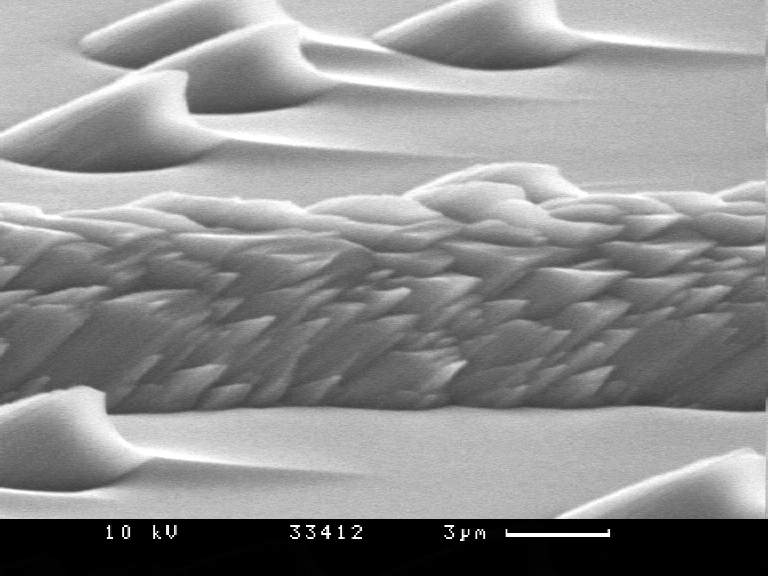
John Bradshaw <joh### [at] nospamhotmailcom> schrieb in im Newsbeitrag:
3c741eae$1@news.povray.org...
> > And what i really miss in these images is the so-called "Kanten Effekt"
> > characteristic to SEM images. In short this means that thin and sharp
> > parts of the object show up much lighter than the more even and smooth
> > parts.
>
> Do you have an example of this effect? Could it be simulated with media?

I have. The sawtooth-like structures in the upper part of the picture get
thinner on the right side. So more secondary electrons emerge from the
sample which brighten up the image in that parts. ("Kante" means "edge", for
those who don't speak german.)

BTW, there _can_ be a "directional lighting" in SEM images. It just depends
on how much voltage you apply to the detector grid. If, for example, the
voltage is zero, you collect only those electrons flying directly into the
detector, leaving the surfaces facing away from it completely dark.

In POV you could perhaps simulate SEM images by radiosity. If you put a thin
horizontal ring around the scene, with ambient=1 all parts facing horizontal
would be the brightest, and all facing vertical would be the darkest,
assumed the camera is looking along -y.

Michael
>
>
Post a reply to this message
|
|
| |
| |
|
|
|
|
| |
| |
|
|
D'oh!
Post a reply to this message
Attachments:
Download '33412_1.JPG' (42 KB)
Preview of image '33412_1.JPG'
|
|
| |
| |
|
|
|
|
| |
| |
|
|
Yowch! This one's scary...

36/700  http://www.uq.edu.au/nanoworld/images_1.html

--
signature{
  "Grey Knight" contact{ email "gre### [at] yahoocom" }
  site_of_week{ url "http://digilander.iol.it/jrgpov" }
}
Post a reply to this message
|
|
| |
| |
|
|
|
|
| |
| |
|
|
In article <3c7431cb$1@news.povray.org>, zie### [at] atlantiswh2tu-dresdende
says...
>
>
> I have. The sawtooth-like structures in the upper part of the picture get
> thinner on the right side. So more secondary electrons emerge from the
> sample which brighten up the image in that parts. ("Kante" means "edge", for
> those who don't speak german.)
>
> BTW, there _can_ be a "directional lighting" in SEM images. It just depends
> on how much voltage you apply to the detector grid. If, for example, the
> voltage is zero, you collect only those electrons flying directly into the
> detector, leaving the surfaces facing away from it completely dark.

True: in that case you collect back-nscattered electrons only.

>
> In POV you could perhaps simulate SEM images by radiosity. If you put a thin
> horizontal ring around the scene, with ambient=1 all parts facing horizontal
> would be the brightest, and all facing vertical would be the darkest,
> assumed the camera is looking along -y.
>
> Michael

--
Regards,
Sander
Post a reply to this message
|
|
| |
| |
|
|
|
|
| |